The Esophagus: Anatomy PART 2
Автор: Din M.D.
Загружено: 2018-02-19
Просмотров: 5710
SUBSCRIBE: https://goo.gl/RHWYGo
LIKE my Facebook page: / dr.din.md
JOIN the Facebook group: https://goo.gl/nx6Tg9
FOLLOW ME on TWITTER: @ DinMD4
FOLLOW ME on INSTAGRAM: din.med.doctor
For EXTRA content including discussion of clinical cases !!
SPECIAL SHOUT-OUT TO:
"The Comical Anatomist": https://thecomicalanatomist.com
Instagram: @thecomicalanatomist
For collaborating with his entertaining image of Lymphatic Drainage of the Esophagus. If you want to see more images like that please follow him and support his work!
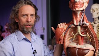
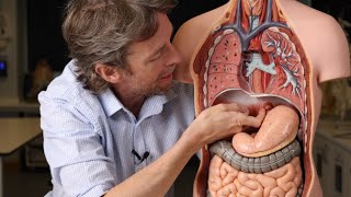
In the Anatomy of the Esophagus PART 2 video, I will talk about the anatomy of the abdominal esophagus, arterial irrigation, venous drainage, lymphatic system and nerves of the esophagus. All the information presented is taken from books or scientific articles. I decided to make it shorter to improve retention, in the next video, we will talk about the clinical application of this video.
Thank you for watching this video! If you enjoyed it please subscribe, like and share if you want me to keep creating more videos. Help me bring medical knowledge to every corner of the world! These videos are specially tailored for Med-students, Doctors, or any Healthcare professional. They could be particularly useful for those going through Med-school or anybody interested in improving their knowledge of Medicine.
I would like to know your opinions. What did you think of the video?
PLEASE IF YOU HAVE ANY QUESTIONS, WRITE IN THE COMMENTS. THE 3 QUESTIONS WITH MORE LIKES WILL BE ANSWERED IN THE FOLLOWING VIDEO !!!
If you have some extra information to give or corrections, please, comment below.
———————————————————————————————————————
Bibliographic References:
1. Boisvert, Rene D, et al. “Bilateral Killian-Jamieson Diverticula: A Case Report and Literature Review.” Canadian Journal of Gastroenterology, vol. 24, no. 3, 2010, pp. 173–174., doi:10.1155/2010/701071.
2. Yazaki, E., and D. Sifrim. “Anatomy and Physiology of the Esophageal Body.” Diseases of the Esophagus, vol. 25, no. 4, 2011, pp. 292–298., doi:10.1111/j.1442-2050.2011.01180.x.
3. Yang, Grace S., et al. “Radiographic and Endoscopic Measurements of Esophageal Length in Pediatric Patients.” Annals of Otology, Rhinology & Laryngology, vol. 114, no. 8, 2005, pp. 587–592., doi:10.1177/000348940511400802.
4. Wemyss-Holden, S. A., et al. “Management of Thoracic Duct Injuries after Oesophagectomy.”British Journal of Surgery, vol. 88, no. 11, 2001, pp. 1442–1448., doi:10.1046/j.0007-1323.2001.01896.x.
5. Oezcelik, Arzu, and Steven R. Demeester. “General Anatomy of the Esophagus.” Thoracic Surgery Clinics, vol. 21, no. 2, 2011, pp. 289–297., doi:10.1016/j.thorsurg.2011.01.003.
6. Cuesta, Miguel A., et al. “A New Concept of the Anatomy of the Thoracic Oesophagus: the Meso-Oesophagus. Observational Study during Thoracoscopic Esophagectomy.”Surgical Endoscopy, vol. 29, no. 9, 2014, pp. 2576–2582., doi:10.1007/s00464-014-3972-1.
7. Lander, Anthony, and Jeremy Newman. “Paediatric Anatomy.” Surgery (Oxford), vol. 28, no. 1, 2010, pp. 11–15., doi:10.1016/j.mpsur.2009.10.009.
8. Mirjalili, S. Ali, et al. “A Reappraisal of Adult Thoracic Surface Anatomy.” Clinical Anatomy, vol. 25, no. 7, 2012, pp. 827–834., doi:10.1002/ca.22091.
9. “Chapter 56: Mediastinum.” Gray's Anatomy: The Anatomical Basis of Clinical Practice, by Susan Standring, 41st ed., Elsevier Ltd, 2016, pp. 986–990.+
10. “Chapter 64: Abdominal oesophagus and stomach.” Gray's Anatomy: The Anatomical Basis of Clinical Practice, by Susan Standring, 41st ed., Elsevier Ltd, 2016, pp. 111–112.
11. Drake, Richard L., et al. “Tórax: Mediastino.” Gray Anatomía Para Estudiantes: Tercera Edición, Elsevier, 2015, pp. 222–225.
12. Drake, Richard L., et al. “Abdomen: Vísceras Abdominales.” Gray Anatomía Para Estudiantes: Tercera Edición, Elsevier, 2015, pp. 310.
13. “Capítulo 2: Abdomen/Esófago.” Moore / Anatomía Con Orientación Clínica, by Keith L. Moore et al., 7th ed., Wolters Kluwer Health, 2013, pp. 282–285.
———————————————————————————————————————
Images:
1. Autor: OpenStaxCollege ce https://cnx.org/contents/[email protected]...
2. Autor: "The Comical Anatomist"
https://thecomicalanatomist.com/

Доступные форматы для скачивания:
Скачать видео mp4
-
Информация по загрузке: