Acetabular Fracture Radiographic Evaluation - Everything You Need To Know - Dr. Nabil Ebraheim
Автор: nabil ebraheim
Загружено: 2018-04-03
Просмотров: 79167
Dr. Ebraheim’s educational animated video describes radiographic evaluation of acetabular fractures.
Follow me on twitter:
https://twitter.com/#!/DrEbraheim_UTMC
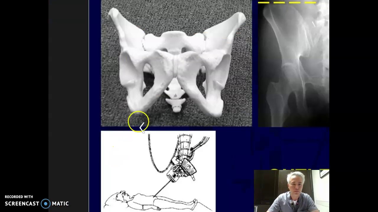
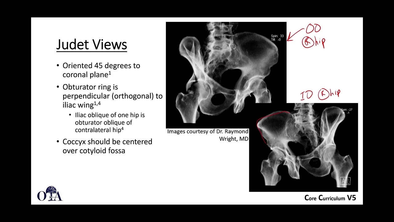
Acetabular fractures usually are evaluated with x-rays and CT scans. These x-rays are usually obtained out of traction. The iliopectineal line represents the anterior column line. The ilioischial line represents the posterior column line. The roof of the acetabulum can be seen in the AP view. The teardrop can be seen in the AP view. The anterior wall of the acetabulum can be seen in the AP view. The posterior wall of the acetabulum can also be seen in the AP view. Lets starts with the obturator view. In the obturator view, you can fully see the obturator foramen. The injured side will be up 45 degrees. Oblique views usually show you a column and opposite wall. In the obturator view, you will see the anterior column and the posterior wall. You can also see the “spur sign.” The spur sign is pathognomonic for both associated BOTH column fractures. In the iliac view, the injured side is down 45 degrees and the good side is up. You will fully see the iliac wing. The posterior column and the anterior wall can be seen in the iliac view. It will tell us how to approach the injury and what type of fracture we have so that we can best approach the injury. CT scan will also show the joint congruity, the size of the fragment, if there is any trapped or impacted fragment. In general, the acetabular wall fracture is oblique and the column fracture is coronal. Transverse fracture of the acetabulum is sagittal or vertical. Transverse fracture of the acetabulum is NOT transverse. The rood arc is an angle between the vertical line through the femoral head and a line through the fracture site on all three views. The question is: did the fracture violate the weight-bearing dome of the acetabulum? You need to know whether you can treat the patient conservatively or surgically. If the fracture does violate the weight-bearing dome, then the patient will be treated surgically. Fracture of the acetabulum that does not violate the weight-bearing dome could be treated conservatively. The problem is that this measurement does not apply to the posterior wall or to the associated both column fractures. The fracture is stable if the fracture line exits outside the weight-bearing dome of the acetabulum and the fracture is usually greater than 45 degrees in the three views (AP, obturator oblique, and iliac oblique). 45 degrees is controversial! Dynamic stress fluoroscopy is used to evaluate the joint stability after an isolated small posterior wall fracture of the acetabulum. It is also used to test the stability of the fracture in a nondisplaced column fracture. If the fracture fragment is more than 50%, then the hip is definitely unstable. When there is a question about the stability of the hip, you should do examination of the hip under anesthesia, regardless of the size of the fracture fragment. The size of the fracture fragment is not a reliable indicator for hip stability. Even if the fracture fragment is less than 20%, you should still do the test. Get the C-arm, use AP and obturator views, flex the hip to 90 degrees and add axial force, and check the hip congruity and the subluxation of the hip. Check for opening of the medial clear space, which indicates instability of the posterior wall.

Доступные форматы для скачивания:
Скачать видео mp4
-
Информация по загрузке: