Скачать
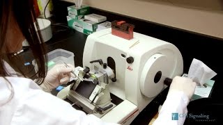
Postbac Life: Lindsey Jay Demonstrates the Microtome
Автор: NIH IRP (Intramural Research Program at the National Institutes of Health)
Загружено: 2019-01-24
Просмотров: 36480
Описание:
https://irp.nih.gov/blog/post/2019/01...
Развернуть
Доступные форматы для скачивания:
Похожие видео
array(20) {
["MRIQZPCX45I"]=>
object(stdClass)#5350 (5) {
["video_id"]=>
int(9999999)
["related_video_id"]=>
string(11) "MRIQZPCX45I"
["related_video_title"]=>
string(25) "Introduction to microtomy"
["posted_time"]=>
string(19) "6 лет назад"
["channelName"]=>
NULL
}
["srICQRjefhU"]=>
object(stdClass)#5360 (5) {
["video_id"]=>
int(9999999)
["related_video_id"]=>
string(11) "srICQRjefhU"
["related_video_title"]=>
string(75) "Основы гистологической техники. Лекция 2."
["posted_time"]=>
string(19) "5 лет назад"
["channelName"]=>
NULL
}
["uEy_NGDfo_8"]=>
object(stdClass)#5348 (5) {
["video_id"]=>
int(9999999)
["related_video_id"]=>
string(11) "uEy_NGDfo_8"
["related_video_title"]=>
string(46) "Using a Micropipette - University of Leicester"
["posted_time"]=>
string(20) "16 лет назад"
["channelName"]=>
NULL
}
["ms3AYSSPa4w"]=>
object(stdClass)#5358 (5) {
["video_id"]=>
int(9999999)
["related_video_id"]=>
string(11) "ms3AYSSPa4w"
["related_video_title"]=>
string(93) "Проверяем работу балансировочных гранул для колес"
["posted_time"]=>
string(25) "2 месяца назад"
["channelName"]=>
NULL
}
["3PBoHU_pidQ"]=>
object(stdClass)#5342 (5) {
["video_id"]=>
int(9999999)
["related_video_id"]=>
string(11) "3PBoHU_pidQ"
["related_video_title"]=>
string(48) "AIDPATH - HISTOLOGICAL TISSUE SAMPLE PREPARATION"
["posted_time"]=>
string(19) "9 лет назад"
["channelName"]=>
NULL
}
["W1F-za1AXFg"]=>
object(stdClass)#5353 (5) {
["video_id"]=>
int(9999999)
["related_video_id"]=>
string(11) "W1F-za1AXFg"
["related_video_title"]=>
string(41) "Leica TP1020 Tissue Processor Programming"
["posted_time"]=>
string(19) "9 лет назад"
["channelName"]=>
NULL
}
["5afOen2AVwg"]=>
object(stdClass)#5346 (5) {
["video_id"]=>
int(9999999)
["related_video_id"]=>
string(11) "5afOen2AVwg"
["related_video_title"]=>
string(58) "Frozen section tutorial -- Embedding and cutting specimens"
["posted_time"]=>
string(20) "12 лет назад"
["channelName"]=>
NULL
}
["hNdCZ_kaDek"]=>
object(stdClass)#5357 (5) {
["video_id"]=>
int(9999999)
["related_video_id"]=>
string(11) "hNdCZ_kaDek"
["related_video_title"]=>
string(30) "Histopathology laboratory Tour"
["posted_time"]=>
string(21) "4 года назад"
["channelName"]=>
NULL
}
["4DJm4NLECQs"]=>
object(stdClass)#5336 (5) {
["video_id"]=>
int(9999999)
["related_video_id"]=>
string(11) "4DJm4NLECQs"
["related_video_title"]=>
string(34) "Histology Techniques and Equipment"
["posted_time"]=>
string(19) "6 лет назад"
["channelName"]=>
NULL
}
["FUddCGVr0bY"]=>
object(stdClass)#5354 (5) {
["video_id"]=>
int(9999999)
["related_video_id"]=>
string(11) "FUddCGVr0bY"
["related_video_title"]=>
string(48) "Wood Sample Preparation for Microscopic Analysis"
["posted_time"]=>
string(20) "10 лет назад"
["channelName"]=>
NULL
}
["N06DiOD37w4"]=>
object(stdClass)#5349 (5) {
["video_id"]=>
int(9999999)
["related_video_id"]=>
string(11) "N06DiOD37w4"
["related_video_title"]=>
string(30) "Histology - Microtome Training"
["posted_time"]=>
string(21) "4 года назад"
["channelName"]=>
NULL
}
["XMjGZHEG4cY"]=>
object(stdClass)#5355 (5) {
["video_id"]=>
int(9999999)
["related_video_id"]=>
string(11) "XMjGZHEG4cY"
["related_video_title"]=>
string(67) "Immunohistochemistry Protocol for Paraffin embedded Tissue Sections"
["posted_time"]=>
string(19) "9 лет назад"
["channelName"]=>
NULL
}
["wvZewru_kF4"]=>
object(stdClass)#5343 (5) {
["video_id"]=>
int(9999999)
["related_video_id"]=>
string(11) "wvZewru_kF4"
["related_video_title"]=>
string(51) "SLEE Automated microtome for MLS Histopathology Lab"
["posted_time"]=>
string(19) "5 лет назад"
["channelName"]=>
NULL
}
["ml4fBEmH8Sg"]=>
object(stdClass)#5341 (5) {
["video_id"]=>
int(9999999)
["related_video_id"]=>
string(11) "ml4fBEmH8Sg"
["related_video_title"]=>
string(29) "Microtome Sectioning Tutorial"
["posted_time"]=>
string(20) "13 лет назад"
["channelName"]=>
NULL
}
["tA7S75MFdNk"]=>
object(stdClass)#5339 (5) {
["video_id"]=>
int(9999999)
["related_video_id"]=>
string(11) "tA7S75MFdNk"
["related_video_title"]=>
string(17) "Cryostat Tutorial"
["posted_time"]=>
string(19) "8 лет назад"
["channelName"]=>
NULL
}
["Oo_O1fZkQqc"]=>
object(stdClass)#5340 (5) {
["video_id"]=>
int(9999999)
["related_video_id"]=>
string(11) "Oo_O1fZkQqc"
["related_video_title"]=>
string(55) "🔬 033 - Different types of microtomes for microscopy"
["posted_time"]=>
string(19) "7 лет назад"
["channelName"]=>
NULL
}
["PFVzBhe-Vnw"]=>
object(stdClass)#5337 (5) {
["video_id"]=>
int(9999999)
["related_video_id"]=>
string(11) "PFVzBhe-Vnw"
["related_video_title"]=>
string(32) "How to Section using a Microtome"
["posted_time"]=>
string(21) "3 года назад"
["channelName"]=>
NULL
}
["x0rzrDU9h0U"]=>
object(stdClass)#5338 (5) {
["video_id"]=>
int(9999999)
["related_video_id"]=>
string(11) "x0rzrDU9h0U"
["related_video_title"]=>
string(41) "Cryosectioning of Fixed and Frozen Tissue"
["posted_time"]=>
string(21) "3 года назад"
["channelName"]=>
NULL
}
["uxtuS1d1wH0"]=>
object(stdClass)#5326 (5) {
["video_id"]=>
int(9999999)
["related_video_id"]=>
string(11) "uxtuS1d1wH0"
["related_video_title"]=>
string(47) "Leica Jung 820 Histocut Rotary Microtome Part 1"
["posted_time"]=>
string(20) "12 лет назад"
["channelName"]=>
NULL
}
["P0cZKCfyUwE"]=>
object(stdClass)#5327 (5) {
["video_id"]=>
int(9999999)
["related_video_id"]=>
string(11) "P0cZKCfyUwE"
["related_video_title"]=>
string(38) "Tissue Processing For Light Microscopy"
["posted_time"]=>
string(20) "10 лет назад"
["channelName"]=>
NULL
}
}
Introduction to microtomy
Основы гистологической техники. Лекция 2.
Using a Micropipette - University of Leicester
Проверяем работу балансировочных гранул для колес
AIDPATH - HISTOLOGICAL TISSUE SAMPLE PREPARATION
Leica TP1020 Tissue Processor Programming
Frozen section tutorial -- Embedding and cutting specimens
Histopathology laboratory Tour
Histology Techniques and Equipment
Wood Sample Preparation for Microscopic Analysis
Histology - Microtome Training
Immunohistochemistry Protocol for Paraffin embedded Tissue Sections
SLEE Automated microtome for MLS Histopathology Lab
Microtome Sectioning Tutorial
Cryostat Tutorial
🔬 033 - Different types of microtomes for microscopy
How to Section using a Microtome
Cryosectioning of Fixed and Frozen Tissue
Leica Jung 820 Histocut Rotary Microtome Part 1
Tissue Processing For Light Microscopy