Broken Instrument Removal - Animation & Clinical Op Sequence: Advanced Endodontics
Автор: endoruddle
Загружено: 2019-06-12
Просмотров: 235386
All content has been transcribed from original audio recordings. As a result, transcribed text may contain certain grammatical, spelling, or other language artifacts which don’t reflect the original audio recording. Please refer to the audio for the most accurate information and messaging. Any reproduction of the video content, audio, or transcribed text is strictly forbidden.
https://www.endoruddle.com/jit/detail...
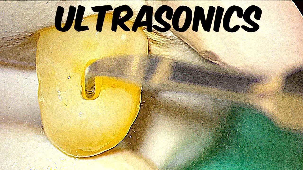
Every clinician who has performed endodontics has experienced a variety of emotions ranging from the thrill-of-the-fill to an upset like the procedural accident of breaking an instrument. When removing a broken instrument, the first option is to utilize piezoelectric ultrasonic technology and specific ultrasonic instruments.
In this Just-In-Time segment, Dr. Ruddle will present via animation and clinical procedure the steps necessary for successful broken instrument removal utilizing ultrasonics. Topics include:
Factors influencing removal
Establishing safe radicular access
Creating a staging platform
Utilizing ultrasonics
Maintaining continuous vision
*
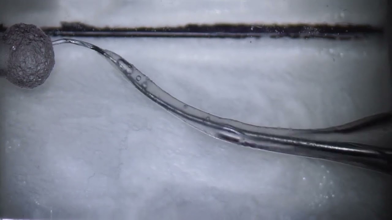
everybody who has practiced endodontics has experienced an upset like breaking an instrument when we break an instrument it's important to assess well angulated pre-operative films and understand the factors that will influence its removal we need to carefully note the position of the broken instrument as it relates from the occlusal table to the apical aspect of the root system we need to carefully take different horizontally angulated films so we can better appreciate their root bulk and form and understand the depth of external root concavities certainly any portion of the instrument that lies in the straight way portion of the canal makes it intriguing whether we can remove the instrument set another way if the entire segment of the broken instrument lies completely around the curvature we won't have a very good opportunity to get direct vision and facilitate the removal efforts clearly most of us that have been removing broken instruments for many years have understood that stainless steel broken instruments are quite easy to remove however in the advent of nickel titanium around 1992 and 1993 has led to an explosion of broken instruments and this material when it's vibrate against with ultrasonic heat presumably causes these instruments to refracture and we may have pieces of the broken segment come out one of the things we've learned from the past is to make a careful radicular access set another way the access should be no bigger than it would have otherwise been if there were no broken instrument in my experience the best tools to create radicular access are a set of gates glidden drills there's two caveats run the gates clintons between 500 and 750 rpm this prevents them from grabbing and being inadvertently sucked into the canal the second suggestion is use them like brushes so we can brush away from Furcal concavities and maximize remain to structure radicular access for me starts with a gates glidden number four whose maximum cross-sectional diameter is 1.1 millimeters this instrument is restricted so that no more than one head depth extends below the orifice the gates glidden 3 follows the 4 it's used one bud depth below the 4 it's working diameter is 0.9 millimeters and this is a classic preparation that has been described in many books across the world over the last 30 to 40 years finally a gates glidden to whose cross-sectional maximum diameter is 7 tenths of a millimeter and this can typically Park right on the head of the broken instrument if you begin to appreciate that each third of the root is about 3 to 5 millimeters then the coronal one-third middle one third and apical one-third come to mind and typically instruments break in the apical 1/3 this means typically around three four or five millimeters of an instrument is usually noticed on a preoperative film it's almost always possible to carry a gates glidden to through the straightaway portions of the canal and park it precisely on the coronal aspect of the broken file well looking ahead we are going to use ultrasonics to try to unwind and back this instrument out but the problem often times clinically is when you praying the ultrasonic instrument down into the root we can't place it's working tip lateral to the broken instrument as such years ago we described how to make a staging platform simply take a transmittal burr or a Jo dandy or any other kind of a cutting instrument and cut the head of the G g2 which was seven tenths of a millimeter maximum height of contour and make a flat run this same number to back into the canal and notice that it creates a little shelf circumferentially around the head of the broken instrument this staging platform will be an optimal way for us to place the appropriately sized ultrasonic instrument the appropriately sized ultrasonic instrument is that instrument that can reach to the clinical

Доступные форматы для скачивания:
Скачать видео mp4
-
Информация по загрузке: