CHAPTER 1 - Anatomy of the Ear: Structural Divisions, Surgical Landmarks, and Embryology
Автор: MED_NESS | MEDICAL BOOKS SIMPLIFIED
Загружено: 2025-12-03
Просмотров: 11
DHINGRA ENT , HEAD & NECK SURGERY
CHAPTER 1
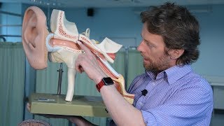
The ear is anatomically segmented into three distinct parts: the external ear, the middle ear, and the internal ear (or labyrinth).
The External Ear This section comprises the pinna, external acoustic canal, and tympanic membrane. The pinna is primarily a framework of elastic cartilage, except for the lobule and the incisura terminalis, a cartilage-free gap between the tragus and crus of the helix utilized for endaural surgical approaches. The external canal is cartilaginous in its outer third and bony in its inner two-thirds, narrowing at the isthmus where foreign bodies frequently become impacted. The canal is lined with skin containing wax-secreting glands in the cartilaginous portion, while the bony portion is lined by thin skin devoid of hair. The nerve supply is complex, involving the vagus (X), trigeminal (V3), facial (VII), and cervical nerves; consequently, Herpes Zoster Oticus manifests with lesions in the concha and posterior tympanic membrane.
The Middle Ear The middle ear cleft is a space lined with mucous membrane that includes the tympanic cavity, eustachian tube, aditus, antrum, and mastoid air cells. The middle ear acts as a six-sided box: the tegmen tympani separates it from the middle cranial fossa superiorly, while the jugular bulb lies inferiorly. The posterior wall houses the pyramid (for the stapedius tendon) and the vertical segment of the facial nerve. A critical surgical landmark here is the facial recess, which allows access to the middle ear without removing the canal wall. The medial wall features the promontory (basal cochlear coil), the oval and round windows, and the facial nerve canal, which can be congenitally dehiscent and prone to injury.
Sound transmission occurs via the ossicular chain (malleus, incus, stapes), while the tensor tympani and stapedius muscles dampen loud sounds to protect the inner ear. The mastoid air cell system connects to the middle ear via the aditus and antrum. During mastoidectomy, surgeons must navigate Korner's septum, a bony plate persisting from the petrosquamosal suture that can obscure deep air cells and the antrum.
The Internal Ear The internal ear consists of a bony labyrinth filled with sodium-rich perilymph and a suspended membranous labyrinth filled with potassium-rich endolymph. The bony labyrinth comprises the vestibule, semicircular canals, and the cochlea, which winds around a central bony pyramid called the modiolus. The membranous labyrinth includes the cochlear duct (hearing), and the utricle, saccule, and semicircular ducts (balance). The utricle and saccule detect linear acceleration via maculae, while the semicircular ducts detect angular acceleration. The endolymphatic sac, located in the subdural space, is the site for drainage surgery in Ménière’s disease. The blood supply is provided exclusively by the labyrinthine artery; as a terminal vessel with no collateral circulation, its obstruction causes permanent ischemic damage.
Embryology The ear develops from three different germ layers: the external canal from the first branchial cleft (ectoderm), the middle ear from the tubotympanic recess (endoderm), and the inner ear from the otic vesicle (ectoderm). Because the inner ear develops independently and reaches maturity by the 20th week, a fetus may possess a functional inner ear even if the external or middle ear is malformed.
--------------------------------------------------------------------------------
Analogy: You can think of the ear as a high-security data center. The External Ear is the satellite dish on the roof, designed to capture signals (sound waves) and funnel them inward. The Middle Ear acts as the transformer room; it takes the raw air signals and mechanically amplifies them (via the ossicles) to travel through a denser medium, much like stepping up voltage for transmission. Finally, the Internal Ear is the liquid-cooled server room deep underground. It is strictly isolated, running on its own dedicated power grid (the terminal labyrinthine artery), and floating in specialized fluids (perilymph and endolymph) to process the data into electrical impulses for the brain. Just as the server room is built before the rest of the office building, the inner ear develops fully in the womb long before the outer structures are finalized.

Доступные форматы для скачивания:
Скачать видео mp4
-
Информация по загрузке: