Visualization during DMEK ophthalmic surgery supported by OCT
Автор: Leica Microsystems
Загружено: 2017-04-03
Просмотров: 912
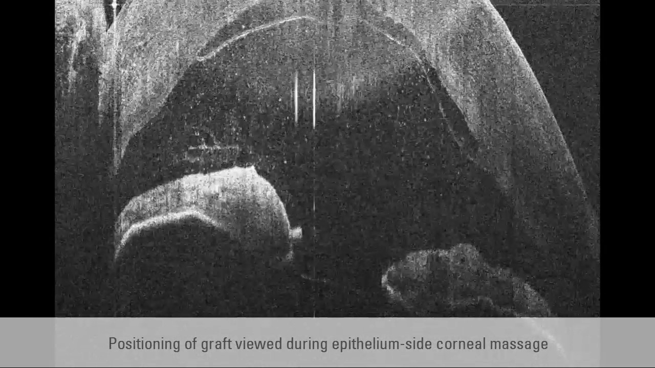
Replacing diseased Descemet’s Membrane with a donor graft is an especially delicate procedure. Accurate positioning and full adherence to the stroma are crucial for optimal patient outcomes. The more visual information the surgeon has of the cornea and the donor and recipient tissues, the more confident he can be of precise graft placement. Watch this video to see how high-resolution intrasurgical optical coherence tomography (OCT) can enhance visualization during DMEK surgery with real-time, cross-sectional imaging of subsurface details.
The video was captured with EnFocus intrasurgical OCT. Learn more about EnFocus: http://bit.ly/2gNlQY5
Read more about optical coherence tomography (OCT) on our Science Lab: http://bit.ly/1PkEj9e
Доступные форматы для скачивания:
Скачать видео mp4
-
Информация по загрузке: