Ultrasound of the Ulnar Nerve in the Cubital Tunnel and its Dynamic Examination
Автор: Dr Kanchan Sharma@Aadhya Pain Management Centre
Загружено: 2024-01-26
Просмотров: 1083
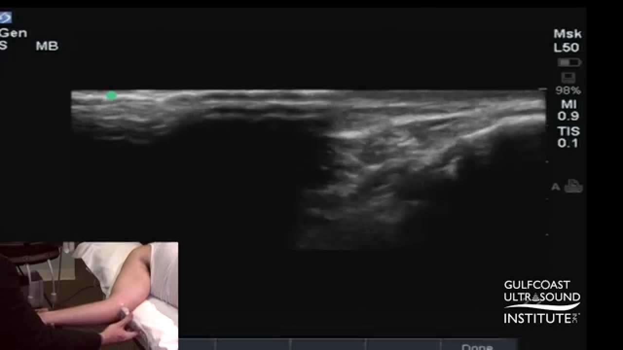
The cubital tunnel, situated on the posterior medial aspect of the elbow, is bordered by the medial epicondyle and olecranon process, with the Cubital retinaculum forming the roof and the posterior band of the ulnar collateral ligament forming the floor. Within this osteo-fibrous passage, the ulnar nerve navigates between the heads of the flexor carpi ulnaris, connected by the arcuate retinaculum. Dynamic examination of the ulnar nerve is crucial for detecting potential subluxation or dislocation. In its normal state, the nerve closely aligns with the medial epicondyle. Any anterior deviation or displacement beyond the common flexor tendon indicates subluxation or dislocation, providing diagnostic insights into cubital tunnel syndrome. Cubital tunnel syndrome, the second most common entrapment neuropathy in the upper limb nerves, involves compression of the ulnar nerve within this anatomical tunnel. Utilizing ultrasound, clinicians can identify potential culprits such as joint osteophytes, lipomas, accessory anconeus muscles, ganglion cysts, and other factors contributing to Cubital tunnel syndrome. Compression or irritation can be detected through palpation, Tinel’s sign, and specific maneuvers like the elbow flexion test, helping diagnose cubital tunnel syndrome. This comprehensive approach aids in both diagnosis and understanding the varied causes of ulnar nerve entrapment in the cubital tunnel.

Доступные форматы для скачивания:
Скачать видео mp4
-
Информация по загрузке: