Basic Anatomy Of The Patella - Everything You Need To Know - Dr. Nabil Ebraheim
Автор: nabil ebraheim
Загружено: 2017-09-26
Просмотров: 151724
Dr. Ebraheim's animated educational video describing the basic anatomy of the patella.
The patella is the largest sesamoid bone in the body. The patellar tendon attaches the patella to the top of the tibia. The quadriceps muscle is attached superiorly to the patella. A small part of the quadriceps tendon then continues over the front of the patella to become the patellar tendon. The apex of the patella, which is the distal part is nonarticular. This area does give attachment to the patellar tendon.
Several bursae is seen around the patella:
1-Suprapatellar
2-Prepatellar
3-Infrapatellar
The thickness of the patella is about 2.5 cm. The cartilage thickness is about 5 mm in the middle portion (probably the largest thickness of cartilage in the body).
The patella has two articular facets:
a-Medial facet:
•Proper
•Odd: the odd facet articulates with deep flexion of the knee and it is located in the distal medial portion of the patella.
b-Lateral facet: the lateral facet is longer wider, larger and broader.
The two facets are separated by a vertical ridge. The medial facet is smaller (probably half the size of the lateral facet)
The patellar tendon works with the quadriceps to straighten the leg. Diagram showing forces and constraints applied to the patella during its function.the patella increases the moment arm of the quadriceps by moving the muscle insertion away from the joint axis. This will increase the ability of the quadriceps muscle to produce torque around the knee joint. The patella is fully engaged at 40-45 of flexion and the forces at the patellofemoral joint is about 3-5 times the body weight.
When the patient has a displaced patellar fracture or when the patellar tendon or the quadriceps tendon is torn, the patient will not be able to do active extension of the knee.
When the patellar tendon is ruptured, the quadriceps tendon will pull the patella upwards. if the quadriceps tendon is ruptured, the patellar tendon will pull the patella downward.
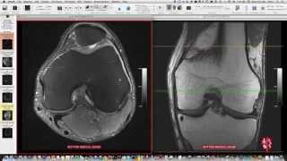
A complete tear of the quadriceps tendon or the patellar tendon can be identified clinically, however it may show on x-ray. MRI may be helpful.
Rupture of the patella tendon causes patella Alta. Rupture of the quadriceps tendon causes patella Baja (patella infera).
The most common problem after a patella fracture fixation is a painful hardware.
The medial patellofemoral ligament inserts at the medial patella at the upper half and it is a restraint to lateral patellofemoral subluxation. Lateral dislocation or subluxation of the patella can be seen in the “sunrise” view. The specific bony bruise pattern may be seen on the medial aspect of the patella and on the lateral femoral condyle (seen on MRI).
Follow me on twitter:
https://twitter.com/#!/DrEbraheim_UTMC
Donate to the University of Toledo Foundation Department of Orthopaedic Surgery Endowed Chair Fund:
https://www.utfoundation.org/foundati...

Доступные форматы для скачивания:
Скачать видео mp4
-
Информация по загрузке: