Osteochondral Lesions Of The Talus - Everything You Need To Know - Dr. Nabil Ebraheim
Автор: nabil ebraheim
Загружено: 2016-12-05
Просмотров: 117244
Dr. Ebraheim’s educational animated video describes Osteochondral Lesions of the Talus.
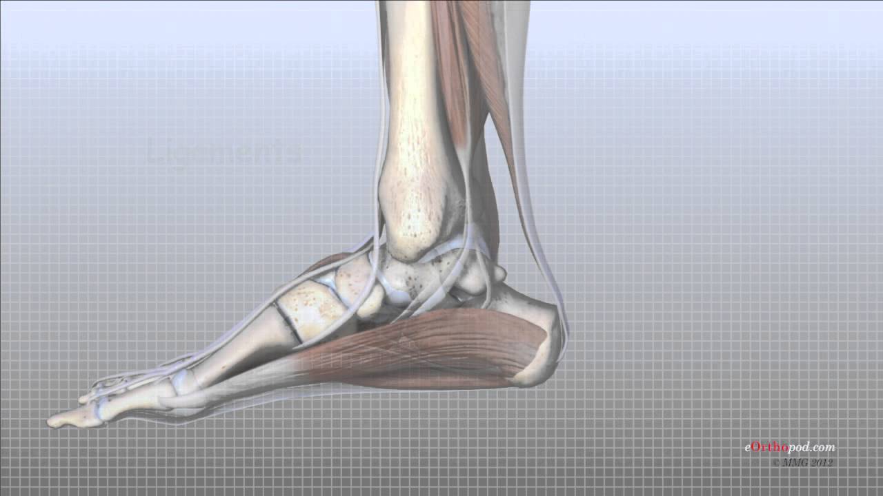
The cartilage thickness of the talus is 1-2 mm, it receives at least 5 times the body weight during normal ambulation, and about 60% of the surface of the talus is covered by cartilage.
It’s not unusual for OCD lesions to occur in the talus.
More than 10% occur bilateral and OCD lesions occur more in males.
About 6
6% of the patients with ankle sprain will have this lesion, and a lot more patients will have the lesion if they have an ankle fracture,
The lesion also occur in patients that are having lateral ligamentous reconstruction.
The lesion may be a contributing cause of the ankle instability.
Most of the patients with symptoms usually play sports and are very active.
Some believe that the primary cause of the OCD lesion is trauma, however this is controversial.
The patient may have an acute fracture, repeated micro trauma, or the patient may not have a history of trauma at all.
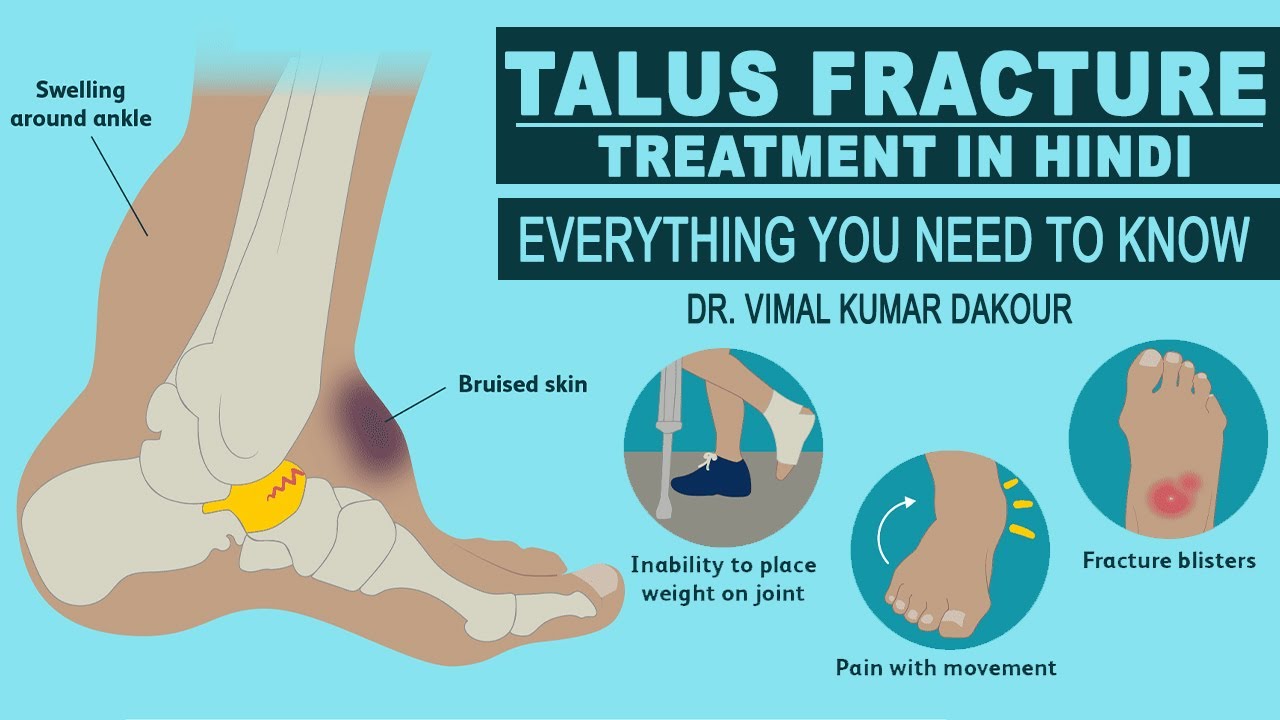
The patient present with either acute inversion injury or chronic ankle pain with swelling, catching, locking and possible ankle instability.
When the patient complains of frequent instability of the ankle, the evidence of ligament laxity on stress view x0ray are usually absent.
Most of the patient who have symptoms are usually active individuals in their 20’s or 30’s.
Get x-ray
Beside x-rays, there are other studies that should be taken.
You can get an MRI which is the study of choice if you suspect the lesion.
When you treat the ankle sprain and it does not get better, you want to rule out an occult lesion of the talus.
A fluid signal behind the lesion on MRI indicates that there is a continuation between the joint and the lesion; the lesion could be unstable and less likely to heal by itself.
You can get a CT scan which is the study of choice if you know there is a lesion and you want to follow that lesion.
It is interesting how the x-ray staging and the MRI staging are almost close in comparison.
x-ray staging:
1- Subchondral compression fracture.
2- Partial detachment of the fracture.
3- Complete detachment of the fracture with no displacement.
4- Complete detachment of the fracture with complete displacement (free fragment).
MRI staging:
1- Articular cartilage edema.
2- Fracture similar to the x-ray.
3- Fracture that is detached but not displaced.
4- Completely displaced fracture fragment. (Stage III and stage IV appear the same as in x-ray stage).
5- Subchondral cyst formation.
It should be noted that the radiographic and the arthroscopic findings do not always correlate.
What are the lesions that you will usually see?
Two types of lesions:
• Posteromedial
• Anterolateral
I find that these lesions are actually opposite to common sense understanding.
The medial lesions:
• Common
• Usually non traumatic
• Larger /deeper
• Posterior
The medial lesions are less symptomatic.
The lateral lesions:
• Less common
• Traumatic
• Smaller/ shallow
• Anterior or slightly central
The lateral lesion is usually symptomatic and difficult to treat without surgery, it has a lower incidence of spontaneous healing and becomes displaced I the joint and symptomatic.
Displaces means that the lesion is a stage IV I either the x-ray or MRI classification.
Usually this lateral lesion occurs due to an inversion or inversion dorsiflexion trauma.
Treatment:
You treat this lesion conservative first by non- weight bearing, short leg cast, or a boot for 4-6 weeks especially if the lesion is acute and nondisplaced.
In general, if the lesion is a lower grade lesion such as stage I or stage II, you will treat the session conservatively.
Surgery is done if the conservative treatment fails.
Surgery is also done if the lesion is a high grade lesion such as stage III or stage IV by MRI.
Surgery is usually done arthroscopic.
In general, if the lesion is a lower grade lesion such as stage I or stage II, you will treat the lesion conservatively.
Treatment of lesions stage III and stage IV is surgical.
Non displaced lesions are treated with immobilization and they may heal.
Regardless how the lesion appears on x-rays, if the patient is asymptomatic after conservative treatment then observe the patient even if there is no evidence of healing of the lesion on x-ray or MRI.
Loose fragments on x-rays are indication for surgery.
If it is an acute fracture, especially the anterolateral fragment, consider reduction and fixation with bioabsorbable.
If the OCD lesion is less than 1 cm, then you will do excision, curettage, or drilling of the lesion.
If it was greater than 1 cm with cartilage intact or the cap intact, you will do retrograde drilling or bone graft.
If the lesion is greater than 1 cm and displaced, then you will do open reduction and internal fixation or you will do osteochondral grafting.

Доступные форматы для скачивания:
Скачать видео mp4
-
Информация по загрузке:


![ЛУЧШЕЕ время восстановления перелома лодыжки и сломанной лодыжки 2022 [25 ЛУЧШИХ СОВЕТОВ!]](https://imager.clipsaver.ru/LZvxS101Ly4/max.jpg)