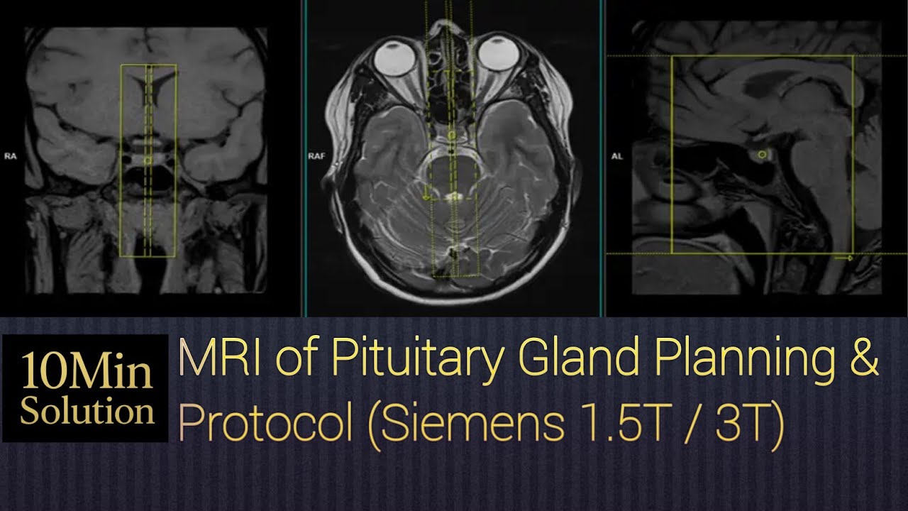
Pituitary gland MRI scan protocols, positioning and positioning
Автор: 10 Min Solution BD
Загружено: 2025-11-06
Просмотров: 132
1. Purpose / Indications
Evaluation of pituitary adenoma (microadenoma or macroadenoma)
Hypopituitarism or hyperprolactinemia
Cushing’s disease, acromegaly, diabetes insipidus
Empty sella, Rathke’s cleft cyst, craniopharyngioma
Post-operative or follow-up imaging
2. Patient Preparation
No special preparation required (unless contrast planned).
Ask about allergies, renal function, and pregnancy.
Remove all metallic items.
Patient should avoid movement during the scan.
3. Patient Positioning
Position: Supine
Head: In a head coil (8-channel or higher preferred)
Centering: At the level of the nasion or mid-sella
Head alignment:
Head should be straight and immobilized.
Align the intercommissural line (AC-PC line) parallel to the table.
Immobilization: Use pads to minimize motion.
4. MRI Protocol (Typical Sequences)
Sequence Plane Slice Thickness Comments
T1-weighted spin echo (SE) Sagittal 2–3 mm For overall anatomy and midline structures
T1-weighted SE Coronal 2–3 mm Best for pituitary stalk and gland morphology
T2-weighted FSE Coronal ± Sagittal 2–3 mm For cystic/edematous lesions
FLAIR (optional) Axial 4 mm For adjacent brain pathology
Dynamic Contrast-Enhanced T1 Coronal 1.5–2 mm Rapid sequential imaging during and after contrast (helps identify microadenomas)
Post-Contrast T1 SE Coronal + Sagittal 2 mm For enhancement pattern and cavernous sinus invasion
3D T1 GRE (optional) Sagittal/Coronal 1 mm isotropic High-resolution reconstruction
5. Contrast Administration
Gadolinium-based contrast: 0.1 mmol/kg (typically 10–15 ml)
Inject at 2 mL/sec with saline flush.
Dynamic sequence begins immediately at the start of injection (to detect differential enhancement between normal gland and microadenoma).
MRI pituitary gland
pituitary MRI protocol
pituitary MRI positioning
MRI sella turcica
dynamic pituitary MRI
MRI brain protocol
MRI microadenoma
radiology tutorial
MRI scan procedure
MRI technologist training
MRI brain and pituitary
MRI planning pituitary
pituitary adenoma MRI
radiology protocol 2025
MRI head coil positioning
7. Imaging Tips
Use thin slices (≤3 mm) for optimal detection of microadenomas.
Dynamic contrast study is key for small lesions.
Include optic chiasm and cavernous sinuses in the field.
Avoid oblique head tilt to maintain true coronal/sagittal planes.
If post-op, include fat-suppressed post-contrast sequences to differentiate scar from recurrence.
8. Typical Total Scan Time
⏱️ Around 20–25 minutes, depending on dynamic protocol timing.
Summary – Key Technical Points
Coil: Head coil
Plane of reference: AC–PC line
Slice thickness: ≤3 mm
Contrast: Mandatory for dynamic study
Dynamic phase timing: 5–6 sets every 10–15 seconds after injection
Доступные форматы для скачивания:
Скачать видео mp4
-
Информация по загрузке:


