Ultrasound Imaging of Morton’s Neuroma | Foot Pain Diagnosis
Автор: SportDrDinesh
Загружено: 2024-10-24
Просмотров: 181
🦶 How to Image a Morton’s Neuroma with Ultrasound
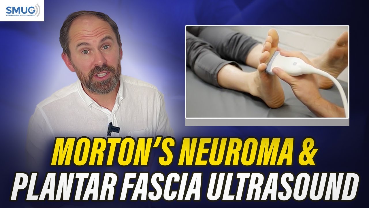
Hi everyone, Dr. Dinesh Sirisena here! In this video, I walk you through the step-by-step process of ultrasound imaging for Morton’s Neuroma, a common cause of foot pain and discomfort, especially between the toes.
Whether you’re a radiologist, sports medicine doctor, sonographer, or clinician learning MSK ultrasound techniques, this tutorial offers valuable insights to improve your diagnostic skills.
🎯 What You’ll Learn:
Ideal patient positioning for comfort and accuracy
Probe placement: short axis vs long axis orientation
How to identify metatarsal heads and web space swelling
Recognizing the hypoechoic appearance of a Morton’s Neuroma-Tips for detecting Mulder’s click and compressible lesions
📸 This video covers real-time ultrasound views and explains what to look out for in both transverse and longitudinal views when diagnosing interdigital neuroma.
🧠 Bonus Tip: Lower the probe frequency for better penetration through thicker plantar skin.
📚 Perfect for clinicians treating:
Forefoot pain
Nerve entrapment in the foot
Plantar discomfort
Sports-related foot injuries
🔍 High-Volume Keywords Covered:
#MortonsNeuroma #FootPain #UltrasoundImaging #MSKUltrasound #NeuromaUltrasound #InterdigitalNeuroma #PlantarFootPain #MusculoskeletalUltrasound #DrDineshSirisena
👍 If you found this useful, please hit Like, Subscribe, and turn on Notifications for more ultrasound tutorials and MSK imaging tips.
🌐 Learn more about my clinical practice: https://www.sportdoctor.com.sg
📲 Follow me on social: @drdineshsirisena (Twitter, Instagram, TikTok)
🎥 Check out my full MSK playlist: [Insert playlist link]
Thanks again for watching, and I hope to see you in the next video!

Доступные форматы для скачивания:
Скачать видео mp4
-
Информация по загрузке: