Популярное

Музыка Кино и Анимация Автомобили Животные Спорт Путешествия Игры Юмор

Интересные видео

2025 Сериалы Трейлеры Новости Как сделать Видеоуроки Diy своими руками

Топ запросов

смотреть а4 schoolboy runaway турецкий сериал смотреть мультфильмы эдисон
dTub
Скачать

Ankle Ligaments Anatomy - Everything You Need To Know - Dr. Nabil Ebraheim

Автор: nabil ebraheim

Загружено: 2013-05-10

Просмотров: 134388

Описание:

Dr. Ebraheim’s educational animated video describes the anatomy of the ankle ligaments.
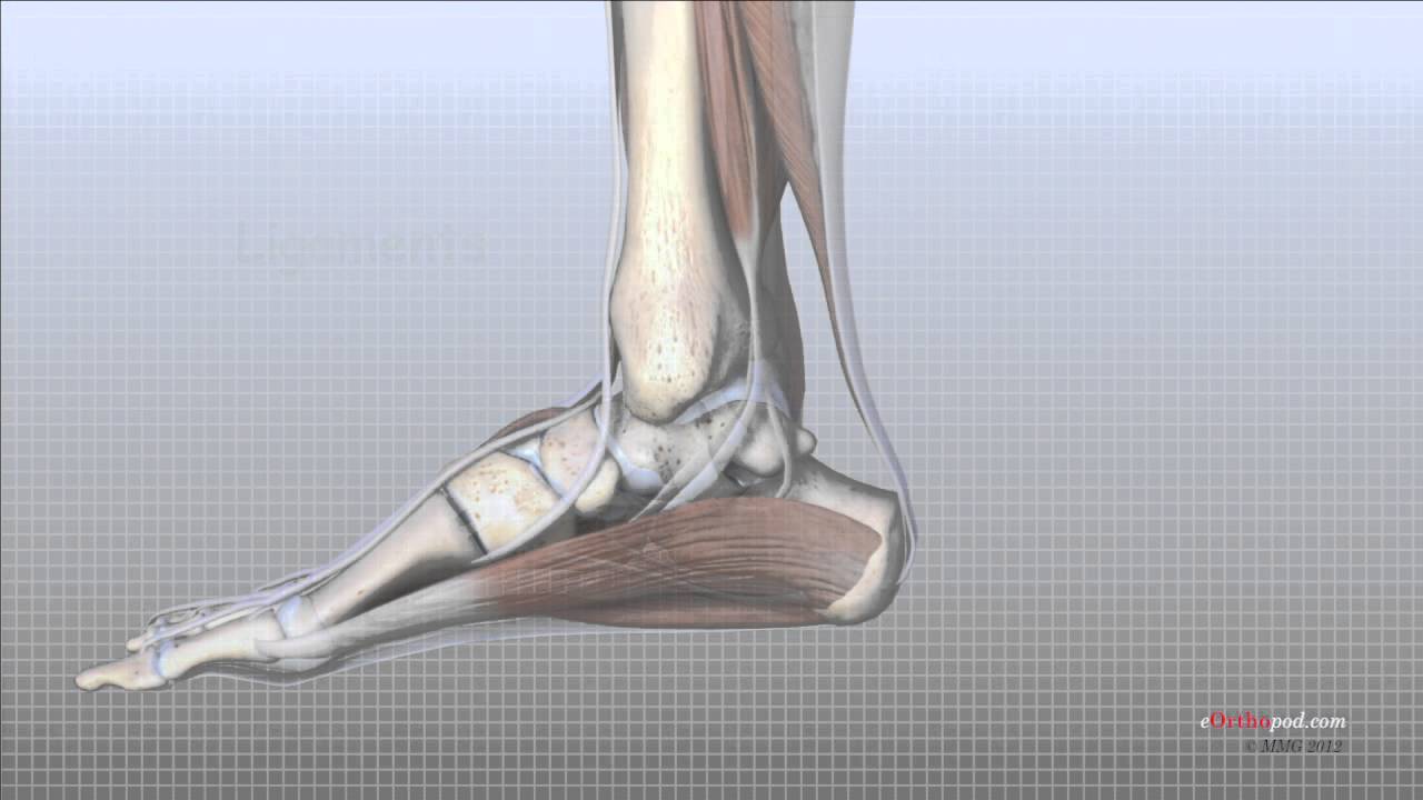
The ankle joint is made of three bones, the tibia, the fibula and the talus. The tibia is the major bone of the lower leg which bears the majority of the body weight. At the angle, the bump of the tibia forms the medial malleolus. The fibula is the smaller of the two bone of the leg. The lateral end of the fibula forms the lateral malleolus. In the ankle joint the talus articulates with the tibia. The talus is involved in multiple movements of the foot.
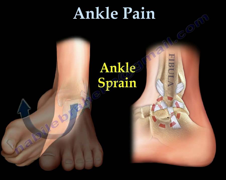
There are ligaments in the ankle that provide connections between the bones. Injury to any of these ligaments may occur when the foot twists, rolls or turns beyond its normal motion. An ankle sprain is a common injury that occurs in sports as basketball and soccer.
The deltoid ligament is on the medial side. It is formed of four parts: anterior tibiotalar part, tibionavicular part, tibiocalcaneal part and the posterior tibiotalar. The superficial deltoid arises from the anterior colliculus. The deep deltoid arises form the posterior colliculus and the intercollicular groove. The deltoid ligament is the main stabilizer of the ankle joint during the stance phase. The deltoid ligament is rarely injured by itself and it is usually associated with fractures.
There are 3 lateral ligaments of the ankle joint:
The anterior talofibular ligament (weakest): origin: 10 mm proximal to the tip of the fibula. Extends from the anterior inferior border of the fibula to the neck of the talus.
The posterior talofibular ligament ( strongest): origin from the posterior border of the fibula. Inserts into posterolateral tubercle of the talus
Calcaneofibular ligament: origin anterior border of the fibula 1 cm proximal to the distal tip. Inserts into the calcaneus distal to the subtalar joint and deep to the peroneal tendon sheath. The lateral ligaments are the most commonly injured ligaments in the ankle.
The ligament of the syndesmosis
•Anterior inferior tibiofibular ligament
•Interosseous ligament
•Posterior inferior tibiofibular ligament
The connection of the tibia and fibula is called the syndesmosis.
High ankle sprain = syndesmosis injury 5-10%. Injury of the ligaments above the ankle.

Become a friend on facebook:
  / drebraheim  

Follow me on twitter:
https://twitter.com/#!/DrEbraheim_UTMC

Ankle Ligaments Anatomy - Everything You Need To Know - Dr. Nabil Ebraheim

Поделиться в:

Доступные форматы для скачивания:

Скачать видео mp4

  • Информация по загрузке:

Скачать аудио mp3

Похожие видео

Анатомия голеностопного сустава | Кости, связки и мышцы

Анатомия голеностопного сустава | Кости, связки и мышцы

Ankle Pain, ankle ligaments sprain - Everything You Need To Know - Dr. Nabil Ebraheim

Ankle Pain, ankle ligaments sprain - Everything You Need To Know - Dr. Nabil Ebraheim

MRI Anatomy of Ankle Ligaments: Syndesmosis

MRI Anatomy of Ankle Ligaments: Syndesmosis

Объяснение функции движения голеностопного и подтаранного суставов. Биомеханика стопы — пронация ...

Объяснение функции движения голеностопного и подтаранного суставов. Биомеханика стопы — пронация ...

Talus Fractures - Everything You Need To Know - Dr. Nabil Ebraheim

Talus Fractures - Everything You Need To Know - Dr. Nabil Ebraheim

Не можешь проплыть 15 минут без остановки и быстро устаешь? Смотри это видео!

Не можешь проплыть 15 минут без остановки и быстро устаешь? Смотри это видео!

5 операций, которые я, как врач, НИКОГДА бы не сделал! / Вы ПОЖАЛЕЕТЕ об ЭТИХ операциях!

5 операций, которые я, как врач, НИКОГДА бы не сделал! / Вы ПОЖАЛЕЕТЕ об ЭТИХ операциях!

Анатомия связок голеностопного сустава | Обзор эксперта-физиотерапевта

Анатомия связок голеностопного сустава | Обзор эксперта-физиотерапевта

Chronic Lateral Ankle Instability - Everything You Need To Know - Dr. Nabil Ebraheim

Chronic Lateral Ankle Instability - Everything You Need To Know - Dr. Nabil Ebraheim

У меня болит лодыжка вот здесь! 11 типичных болевых точек и что они означают

У меня болит лодыжка вот здесь! 11 типичных болевых точек и что они означают

Ankle MRI anatomy

Ankle MRI anatomy

Subtalar Arthroereisis ProStop®

Subtalar Arthroereisis ProStop®

Кости стопы и голеностопного сустава | Подготовка к рентгенологической анатомии, часть 1 | Анатом...

Кости стопы и голеностопного сустава | Подготовка к рентгенологической анатомии, часть 1 | Анатом...

Нервы стопы

Нервы стопы

Как укрепить хрящ?

Как укрепить хрящ?

MRI Anatomy of Ankle Ligaments: Deltoid Ligament

MRI Anatomy of Ankle Ligaments: Deltoid Ligament

Боль в лодыжке — всё, что вам нужно знать — доктор Набиль Эбрахайм

Боль в лодыжке — всё, что вам нужно знать — доктор Набиль Эбрахайм

Foot Anatomy Animated Tutorial

Foot Anatomy Animated Tutorial

Échographie de la cheville

Échographie de la cheville

Анатомия бедренной грыжи

Анатомия бедренной грыжи

© 2025 dtub. Все права защищены.


  • Контакты
  • О нас
  • Политика конфиденциальности


Контакты для правообладателей: infodtube@gmail.com