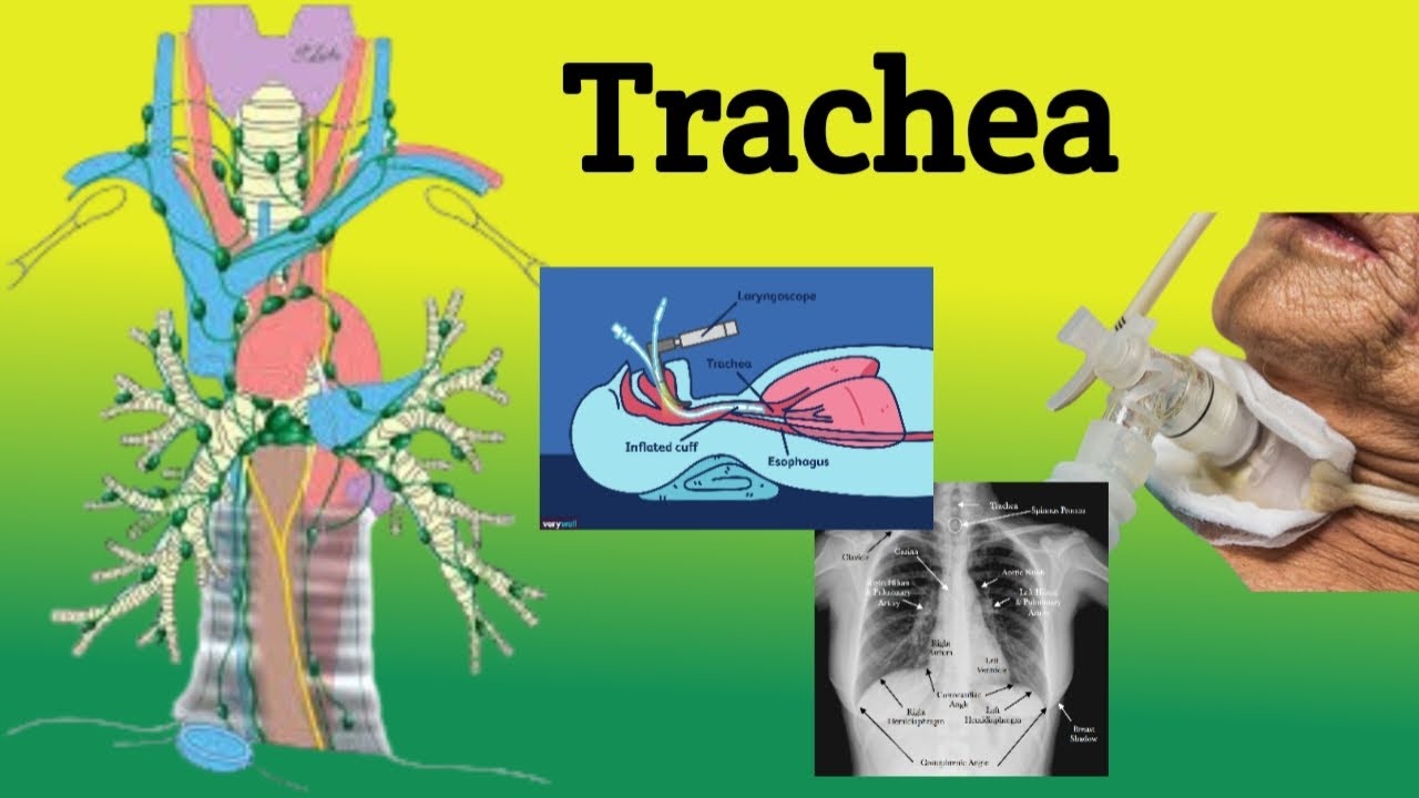
Trachea || Anatomy; Location, Relations, Clinical Correlations
Автор: Let's make medicine easy
Загружено: 2024-07-14
Просмотров: 492
Anatomy of the Trachea
Location and Relations
***The Trachea in the Neck*** 🌟
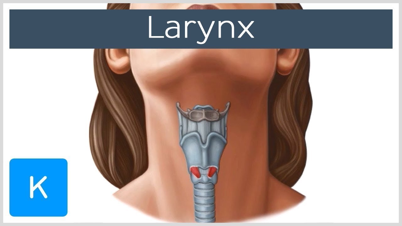
The trachea begins at the inferior margin of the cricoid cartilage (C6 vertebral level) and extends inferiorly into the superior mediastinum.
In the neck, the trachea is covered anteriorly by the isthmus of the thyroid gland, the inferior thyroid veins, the sternothyroid and sternohyoid muscles, and the cervical fascia.
Laterally in the neck, the trachea is related to the common carotid arteries, the thyroid gland lobes, the inferior thyroid arteries, and the recurrent laryngeal nerves.
Posteriorly in the neck, the trachea is in contact with the esophagus and the vertebral column.
***The Trachea in the Thorax*** 🌟
In the thorax, the trachea lies in the superior medi
Anatomy of the Trachea
Location and Relations
*The Trachea in the Neck* 🌟
The trachea begins at the inferior margin of the cricoid cartilage (C6 vertebral level) and extends inferiorly into the superior mediastinum.
In the neck, the trachea is covered anteriorly by the isthmus of the thyroid gland, the inferior thyroid veins, the sternothyroid and sternohyoid muscles, and the cervical fascia.
Laterally in the neck, the trachea is related to the common carotid arteries, the thyroid gland lobes, the inferior thyroid arteries, and the recurrent laryngeal nerves.
Posteriorly in the neck, the trachea is in contact with the esophagus and the vertebral column.
*The Trachea in the Thorax* 🌟
In the thorax, the trachea lies in the superior mediastinum.
Anteriorly, the trachea is covered by the manubrium of the sternum, the remains of the thymus, the left brachiocephalic vein, the aortic arch, the brachiocephalic trunk, the left common carotid artery, and the deep cardiac plexus.
On the right side, the trachea is related to the pleura, the right vagus nerve, and the brachiocephalic trunk near the root of the neck.
On the left side, the trachea is related to the left recurrent laryngeal nerve, the aortic arch, and the left common carotid and subclavian arteries.
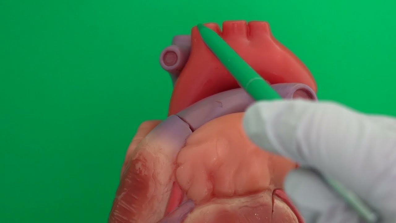
The trachea bifurcates into the right and left main bronchi at the level of the sternal angle (T4-T5 vertebral level), forming the carina.
Structure 🔬
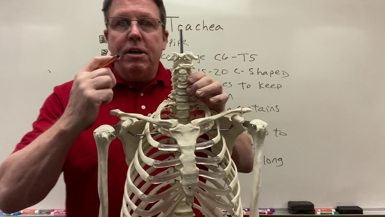
The trachea is a tube-like structure composed of 16-20 C-shaped cartilage rings that occupy the anterior and lateral aspects of the tracheal wall.
The posterior aspect of the trachea is a flat, membranous wall containing the trachealis muscle (smooth muscle).
The tracheal lumen is lined by a mucosa of pseudostratified ciliated columnar epithelium with goblet cells.
The tracheal cartilage rings are connected by fibroelastic tracheal annular ligaments.
Arterial Supply and Venous Drainage 💉
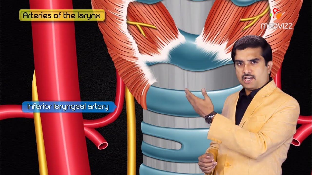
The proximal half of the trachea is supplied by tracheo-esophageal branches of the inferior thyroid artery.
The distal half of the trachea and the carina are supplied by the superior and middle bronchial arteries.
The trachea drains venously into the inferior thyroid venous plexus.
Lymphatic Drainage 💧
The lymphatic drainage of the trachea is to the pretracheal and paratracheal lymph nodes.
Innervation 🧠
The trachea receives sensory innervation from the recurrent laryngeal nerve.
Autonomic innervation is provided by the anterior and posterior pulmonary plexuses, originating from the sympathetic and parasympathetic fibers.
Clinical Correlations 🏥
Tracheal anomalies, such as tracheal bronchus ("pig bronchus"), tracheal diverticulum, and tracheal buckling, can occur and may present with respiratory symptoms.
Tracheal deviation or shift can occur due to mediastinal masses or other pathologies, which can be clinically relevant.
The carina, the ridge of cartilage between the openings of the two bronchi, is the most sensitive area of the trachea for triggering the cough reflex.
References
[1] [Elsevier: Respiratory System - Trachea](https://www.elsevier.com/resources/an...)
[2] [TeachMeAnatomy: Tracheobronchial Tree](https://teachmeanatomy.info/thorax/or...)
[3] [Medscape: Trachea Overview](https://emedicine.medscape.com/articl...)
[4] [Radiopaedia: Trachea](https://radiopaedia.org/articles/trachea)
[5] [ScienceDirect: Trachea](https://www.sciencedirect.com/topics/...)
[6] [NCBI: Trachea](https://www.ncbi.nlm.nih.gov/pmc/arti...)
[7] [Slideshare: Anatomy of Trachea](https://www.slideshare.net/slideshow/...)
Time Chapters
00:00 Introduction
00:50 Location
04:36 Comparative features of adult and child trachea
06:42 Relations in cervical region
10:02 Relations in thoraxic region
13:13 Blood supply
14:14 Nerve supply
14:28 Clinical correlations
Доступные форматы для скачивания:
Скачать видео mp4
-
Информация по загрузке: