Anatomy of Lower Limb Rapid Revision - By Dr. Monalisa🚀
Автор: Dr.G.Bhanu Prakash
Загружено: 2024-09-21
Просмотров: 31600
📌 𝐅𝐨𝐥𝐥𝐨𝐰 𝐨𝐧 𝐈𝐧𝐬𝐭𝐚𝐠𝐫𝐚𝐦:- / drgbhanuprakash
📌𝗝𝗼𝗶𝗻 𝗢𝘂𝗿 𝗧𝗲𝗹𝗲𝗴𝗿𝗮𝗺 𝗖𝗵𝗮𝗻𝗻𝗲𝗹 𝗛𝗲𝗿𝗲:- https://t.me/bhanuprakashdr
Anatomy of Lower Limb Rapid Revision - By Dr. Monalisa🚀
Bones of the Lower Limb🦴
Pelvic Girdle: Comprising the ilium, ischium, and pubis, the pelvic girdle connects the lower limb to the trunk. Understanding the anatomy of the pelvic girdle is crucial for identifying various clinical conditions such as fractures, hip dislocations, and congenital hip dysplasia.
Femur: The longest and strongest bone in the body, the femur is vital for weight-bearing and locomotion. Dr. Monalisa highlights the anatomical features of the femur, including the head, neck, greater and lesser trochanters, and the shaft, focusing on areas prone to fractures.
Patella: Known as the kneecap, the patella protects the knee joint and improves the leverage of the quadriceps muscle. Learn about its anatomical position, articulation, and common pathologies like patellar dislocation and fractures.
Tibia and Fibula: These two bones form the skeleton of the leg. Dr. Monalisa explains the anatomical differences between them, their articulations at the knee and ankle, and clinical conditions such as tibial fractures and compartment syndrome.
Bones of the Foot: The foot consists of tarsals, metatarsals, and phalanges. Understanding the anatomy of the foot is essential for diagnosing common conditions like plantar fasciitis, fractures, and deformities such as flatfoot and clubfoot.
2. Joints of the Lower Limb ⚙️
Hip Joint: A ball-and-socket joint that provides stability and a wide range of motion. Dr. Monalisa covers the structure of the hip joint, including the acetabulum, femoral head, joint capsule, and ligaments, as well as clinical aspects like hip osteoarthritis and dislocation.
Knee Joint: The largest and most complex joint in the body, the knee joint is crucial for movement and stability. This section explores the components of the knee, including the femur, tibia, patella, menisci, and ligaments (ACL, PCL, MCL, LCL). Common pathologies such as meniscal tears, ligament injuries, and patellar tracking disorders are also discussed.
Ankle Joint: A hinge joint formed by the tibia, fibula, and talus, responsible for dorsiflexion and plantarflexion. Dr. Monalisa explains the anatomy of the ankle, including the ligaments that provide stability, and covers common conditions like ankle sprains and fractures.
Joints of the Foot: These include the subtalar joint, midtarsal joint, and metatarsophalangeal joints. Understanding these joints is vital for diagnosing foot pathologies like bunions, hammertoes, and arthritis.
3 Muscles of the Lower Limb💪
Gluteal Region: The gluteal muscles, including the gluteus maximus, medius, and minimus, play a key role in hip movement and stabilization. Dr. Monalisa highlights their attachments, innervations, and functions, as well as their clinical relevance in conditions like hip bursitis and gluteal muscle injuries.
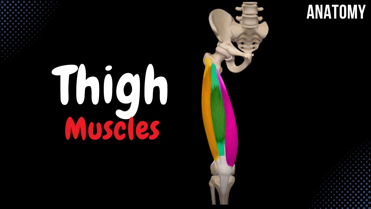
Thigh Muscles: Divided into anterior (quadriceps), posterior (hamstrings), and medial (adductors) compartments, the thigh muscles are essential for movement and support. This section covers their anatomy, functions, and clinical conditions such as quadriceps tendon rupture, hamstring strains, and groin injuries.
Leg Muscles: The leg is divided into anterior, posterior, and lateral compartments. Dr. Monalisa explains the anatomy and functions of key muscles like the gastrocnemius, soleus, tibialis anterior, and fibularis longus, and discusses pathologies such as shin splints and Achilles tendonitis.
Muscles of the Foot: These include intrinsic and extrinsic muscles responsible for toe movements and maintaining the arch of the foot. Learn about the roles of these muscles in foot mechanics and common conditions like plantar fasciitis and flatfoot.
4. Nerves of the Lower Limb🧠
Lumbar and Sacral Plexuses: The lumbar and sacral plexuses give rise to nerves that innervate the lower limb, including the femoral, obturator, sciatic, tibial, and common fibular nerves. Dr. Monalisa provides a detailed overview of these nerves, their courses, branches, and clinical relevance, including nerve injuries and entrapment syndromes.
5. Blood Supply of the Lower Limb🩸
Arterial Supply: The lower limb receives blood from the femoral artery and its branches. Dr. Monalisa discusses the arterial anatomy, highlighting key branches like the profunda femoris, popliteal, anterior tibial, and posterior tibial arteries, and their clinical significance in conditions such as peripheral artery disease.
Venous Drainage: The venous system includes the superficial veins (great and small saphenous veins) and deep veins. Learn about the venous anatomy and its clinical importance in conditions like deep vein thrombosis (DVT) and varicose veins.
#anatomy #anatomylecture #anatomyneetpg #anatomymbbs #anatomyexplained #anatomyusmle
#mbbs

Доступные форматы для скачивания:
Скачать видео mp4
-
Информация по загрузке: