Anatomy Of The Teres Major Muscle - Everything You Need To Know
Автор: nabil ebraheim
Загружено: 2015-04-01
Просмотров: 102632
Dr. Ebraheim’s educational animated video describes the anatomy of the Teres Major muscle.
Origin & insertion: the teres major muscle arises from the dorsal (posterior) aspect of the inferior angle of the scapula and inserts into the medial lip of the intertubercular groove of the humerus.
The teres major muscle is one of several muscles that connect the scapula to the humerus. These muscles include: triceps muscle, infraspinatus muscle, supraspinatus muscle and teres major muscle.
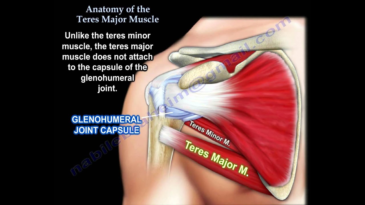
Unlike the teres minor muscle, the teres major muscle does not attach to the capsule of the glenohumeral joint. The teres major muscle has a close insertion into the anterior side of the proximal humerus along with the lattisimus dorsi, pectoralis major and subscapularis muscles.
Innervation: the teres major muscle is innervated by the lower subscapular nerve (C5,C6) of the brachial plexus. The subscapularis muscle is inserted into the humerus higher than the teres major and is innervated by the upper and lower subscapular nerves (C5,C6). The latissimus dorsi muscle is inserted into the humerus between the others and is innervated by the thoracodorsal nerve (C6,C7,C8). The subscapular muscle carries the names subscapular. The teres major muscle is lower than the subscapular muscle so it is innervated by the lower subscapular nerve.
Function: the teres major muscle causes three movements of the shoulder joint:
1-It pulls the humerus towards the trunk (adduction).
2-It turns the humerus medially (internal rotation).
3-It pulls the humerus posteriorly (extension/retroversion).
Important anatomical structures related to the teres major muscle:
1-Arteries: Circumflex scapular artery, posterior circumflex humeral artery, deep brachial artery
2-Nerves: Posterior cord of the brachial plexus, axillary nerve, radial nerve, lower subscapular nerve.
There are three important anatomical spaces located in the posterior shoulder associated with the teres major muscle:
1-Quadrangular space: boundaries:
•teres minor (superior)
•teres major (inferior)
•long head triceps (medial)
•Surgical neck humerus (lateral).
It contains the axillary nerve and the posterior humeral circumflex artery. Axillary nerve can be injured during surgery, fractures, or dislocations. Surgical dissection should be above teres minor muscle to avoid axillary nerve injury.
2-Triangular interval: boundaries:
•Teres major
•Long head triceps
•Humeral shaft
It contains the deep branch of brachial artery and the radial nerve.
3-Triangular space: boundaries:
•Teres major
•Teres minor
•Long head triceps.
It contains the circumflex scapular artery.
Become a friend on facebook:
/ drebraheim
Follow me on twitter:
https://twitter.com/#!/DrEbraheim_UTMC
Donate to the University of Toledo Foundation Department of Orthopaedic Surgery Endowed Chair Fund:
https://www.utfoundation.org/foundati...
Background music provided as a free download from YouTube Audio Library.
Song Title: Every Step
Доступные форматы для скачивания:
Скачать видео mp4
-
Информация по загрузке: